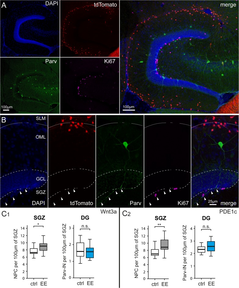
Figure 5.
Environmental enrichment does not affect the densities parvalbumin-expressing interneurons of the dentate gyrus. (A) Identification of parvalbumin-expressing interneurons and proliferating cells in the denate gyrus of a P36 Wnt3a mouse. Top left panel: DAPI counterstaining, top middle panel: tdTomato-labeled cells, bottom left panel: parvalbumin immunohistochemistry, and middle right panel: Ki67 expression in the subgranular zone. The right panel shows all previous images superimposed. Please notice the much larger number of tdTomato expressing Cajal–Retzius cells compared with parvalbumin-labeled interneurons. (B) Left to right: identification at higher magnification of dentate gyrus structure (DAPI counterstaining), Cajal–Retzius cells (tdTomato fluorescence), parvalbumin-positive interneurons (Parv), dividing cells (Ki67), and superimposition of all the images (merge). Arrowheads indicate the main axonal trunk of the interneuron coasting the subgranular zone. (C1) Summary plots showing the densities of Ki-67-positive nuclei of neural progenitor cells (NPC) (left panel) and of parvalbumin immunoreactive cells in Wnt3a mice housed in control (ctrl) and environmental enriched (EE) conditions (left panel). Notice the selective effect of environmental enrichment on neural progenitors compared with interneurons. (C2) Same analysis as in (C1) carried out in sections from PDE1c mice.

