Figure 8.
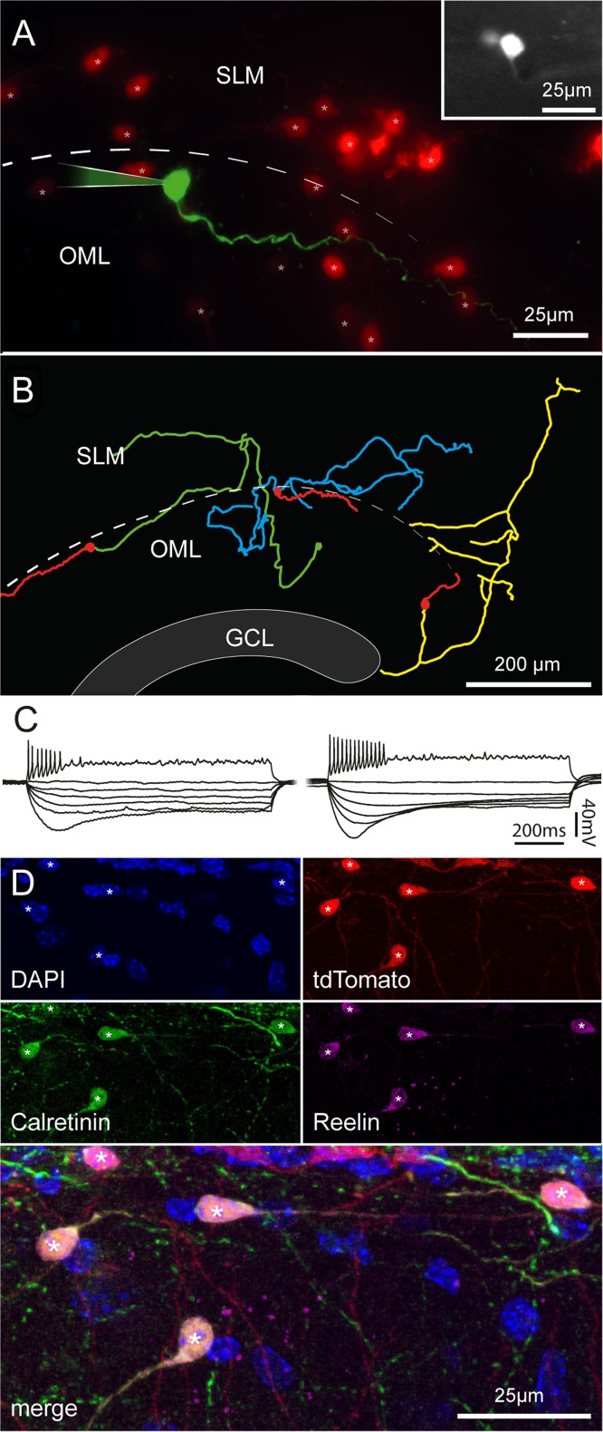
Morphofunctional properties and immunoreactivity of Cajal–Retzius cells in fully mature mice. (A) Pseudocolor image of a biocytin-filled cell (green) form a P153 Wnt3a animal housed under enriched conditions. Other tdTomato-labeled cells are shown in red. Notice the typical tadpole morphology. Inset at the top right shows a fluorescent image of the same cell in the living slice. SLM, stratum lacunosum-moleculare; OML, outer molecular layer, dotted line: hippocampal fissure. (B) Morphological reconstruction of 3 Cajal–Retzius cells from adult mice (Wnt3a, enriched conditions from left to right: P153, P150, and P150). Notice the typical distribution pattern of dendrites and axons. Dendrites: red, axons: green, blue, and yellow. GCL, granule cell layer. (C) Firing patterns and membrane properties recorded in Cajal–Retzius cells of adult mice (P150–P177). Notice the similarity with what recorded in younger animals shown in Figure 2. (D) Cajal–Retzius cells from mature animals express calretinin and reelin. Images from a P112 Wnt3a mouse (housed in control conditions) showing DAPI nuclear counterstaining and labeling of tdTomato, calretinin, and reelin in single pictures, and superimposed. Notice the typical tadpole-like morphology of the cells identified by asterisks. Compare to Figure 2.

