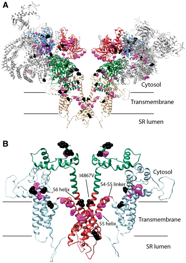
Figure 4.

(A) Homology model of human RyR2. The protein is shown in cartoon form, with the disease hotspots highlighted in colours (hotspot 1: blue, hotspot 2: red; hotspot 3: green; hotspot 4: light blue). View is from the ‘side,’ parallel to the membrane. Only two out of four subunits are shown for clarity. Positions for CPVT-associated variants are highlighted, with atoms shown in Van der Waals representation (purple: associated with cardiac arrest, black: all others). (B) Close-up of the transmembrane region of the RyR2. This region forms the bulk of disease hotspot 4. The following subregions are highlighted: pore-forming region (red), additional transmembrane (cyan), and C-terminal cytosolic extension to the pore (green). Positions of CPVT-associated variants associated with cardiac arrest are highlighted in purple, and all others in black. The S5 and S6 helices and the S4–S5 linker are labelled.
