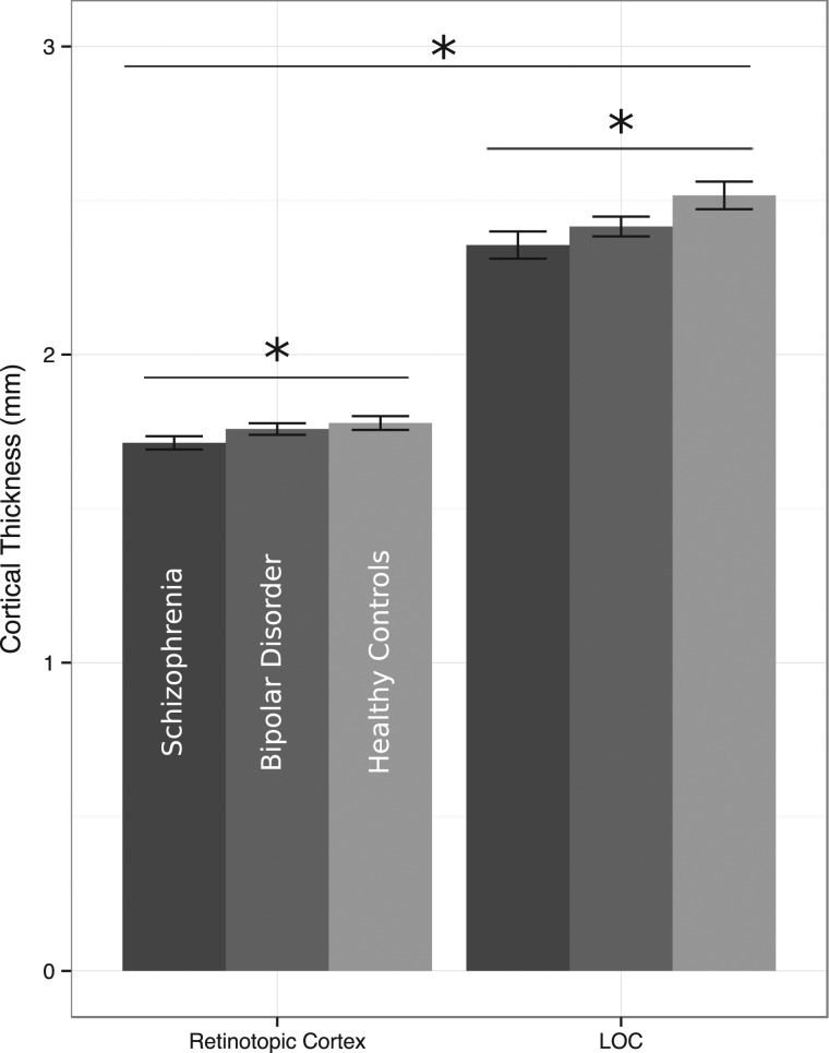
Figure 2.
Comparison of visual ROI cortical thickness by group and region. Across groups, retinotopic cortex was significantly thinner than LOC. Across ROIs, cortical thickness was significantly different between the participant groups: schizophrenia patients had the thinnest cortex in both ROIs, followed by bipolar patients, and healthy controls had the thickest cortex in both areas. In a direct comparison, schizophrenia patients had significantly thinner cortex than healthy controls across both ROIs, but comparisons between bipolar patients and the other two groups showed only trend-level differences. No significant group-by-ROI interaction was present in the data.

