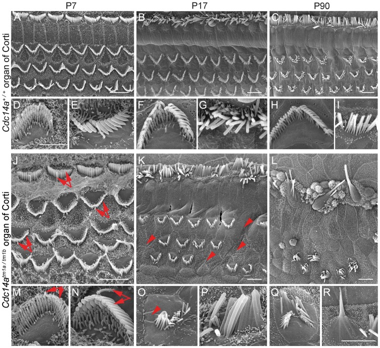
Figure 7.
SEM images of inner (IHC) and outer hair cells (OHC) from the apical turn of the organ of Corti from wild-type and mutant Cdc14a mice showing stereocilia bundle morphology. (A–I) Three rows of OHC and one row of IHC stereocilia from the wild-type at three ages. (J–R) Comparable images from the compound heterozygous Cdc14atm1a/tm1b mouse. Higher magnification views of OHC (D, F, H and M, O, Q) and IHC (E, G, I and N, P, R) stereocilia bundles. IHCs and OHCs of Cdc14atm1a/tm1b mutants at P7 (J, M, N) appear to have normal kinocilia and stereocilia bundle morphology (arrows). At P17, for Cdc14atm1a/tm1b, some stereocilia of IHCs and OHCs fuse and undergo degeneration (K, O, P). Some OHC stereocilia and OHCs themselves are missing (K, O, arrowheads). Extensive degeneration of IHC and OHC stereocilia is observed at P90 (L, Q, R). Many OHC stereocilia bundles are missing (L), and most IHC stereocilia are degenerating (L, R). Scale bars for (A–C) and (J–L) are 5 m. A 2 m scale bar in D applies to D-I and a 2m scale bar in R applies to (M–R).

