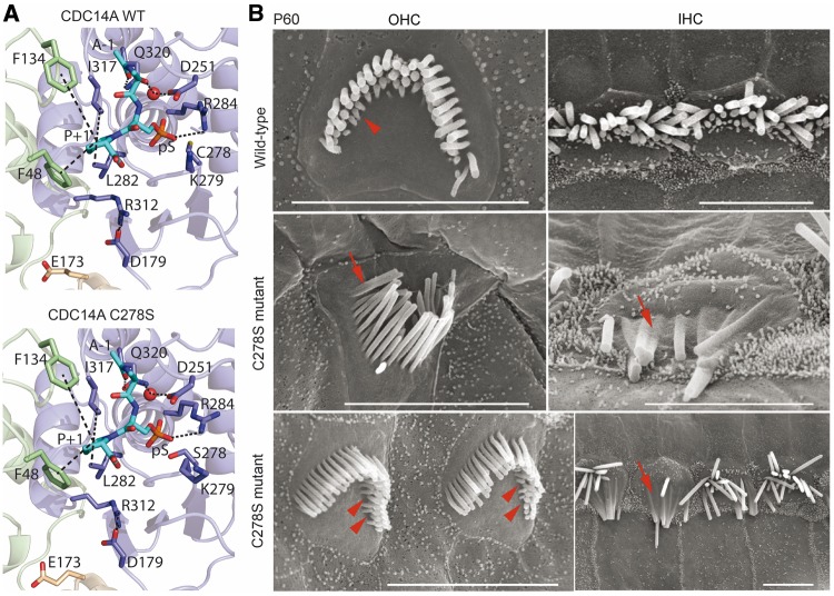
Figure 8.
Structural model of CRISPR/Cas9 edited p.C278S variant of mouse CDC14A protein and SEM images of hair cells. (A) Catalytic site close-up view of structural models of mouse CDC14A wild-type and mutant p.C278S. The substrate (alanine-phosphorylated-serine-proline peptide) is in the binding site shown as cyan sticks where the alanine and the proline are indicated as A-1 and P + 1. The interactions involving the catalytic site residues are indicated by dashes. (B) SEM micrographs of P60 organ of Corti IHC and OHC stereocilia bundles of wild-type (upper row) and a homozygous p.C278S mutant (middle and lower rows). Wild-type OHC stereocilia bundle (upper left) shows three rows of stereocilia. In the shortest third row, most stereocilia are present (arrowhead). Homozygous p.C278S stereocilia are degenerating (middle and lower rows), fusing (arrows), and several third row OHC stereocilia are missing (arrowheads). Scale bars, 5 μm.

