
Figure 1: Adult rats fail to regenerate new hair follicles and new adipocytes after excisional wounding.
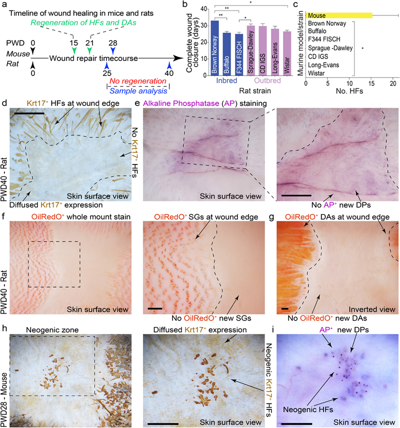
(a) Timeline of full-thickness excisional wound healing in mice and rats. Mouse wounds heal and regenerate new HFs and dermal adipocytes (DA), while rat wounds fail to regenerate. (b) Despite their inability to regenerate, circular (d=2.0 cm) wounds in rats (see Table S1 for biological replicate information) undergo complete re-epithelialization, ranging between 25–33 days depending on the strain (see Table S1 for details). (c) Circular d=2.0 cm wounds in all strains of rats (n=5 per strain) failed to regenerate new HFs and new DA. Wounds in three out of five mixed background mice regenerated new HFs. Of these three mice, average number of regenerated HFs was 15 (see Tables S4 and S4). (d, e) Wholemount Krt17 and alkaline phosphatase (AP) staining revealed lack of new HFs in circular excisional wounds in rats at PWD40. Representative wounds are shown. (f, g) Wholemount OilRedO staining confirms the absence of new HFs (based on the lack of OilRedO+ sebaceous glands (SGs)) and new DA in circular excisional wounds in rats at PWD40. Representative wound is shown. (h, i) Wholemount Krt17 and AP staining reveal new HFs in excisional wounds in mice at PWD28. Representative wounds that underwent HF regeneration are shown. Values in the graphs on 1b and 1c are means ± S.E.M. One-way ANOVA in 1b, P<0.05; Post-hoc Tukey’s multiple comparison test in 1b, *P<0.05, **P<0.01; two-tailed unpaired t-test in 1c, *P=0.0127. Size bars: d-j – 100 µm.