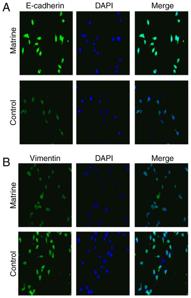
Figure 6.

Effect of matrine on E-cadherin and vimentin expression in Huh-7 cells. Representative single-color and merged images of Huh-7 cells illustrate the immunofluorescence staining for (A) E-cadherin (green) and (B) vimentin (green), with the cell nucleus (blue) stained using DAPI (magnification, ×100). E, epithelial.
