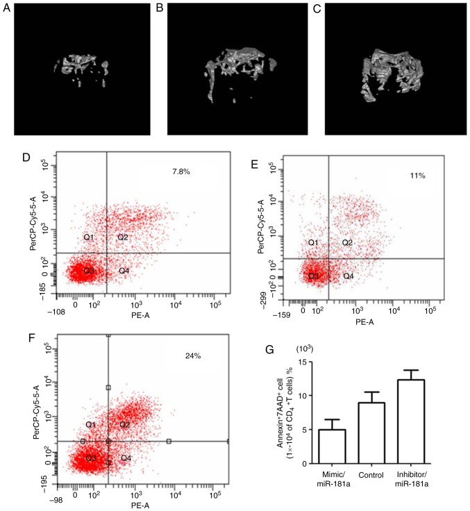
Figure 6.
Representative microtomography images of mouse bone and analysis of CD4+ T lymphocyte apoptosis. Mice were treated with BMMSCs transfected with (A) miR-181a mimic, (B) mimic control or (C) miR-181a inhibitor. Flow cytometric analysis of apoptosis of CD4+T lymphocytes from mice treated with (D) miR-181a mimic, (E) mimic control or (F) miR-181a inhibitor. (G) Comparison of the apoptotic rate of CD4+T lymphocytes in each group. 7AAD, 7-aminoactinomycin D; BMMSCs, bone marrow mesenchymal stem cells; CD4, cluster of differentiation 4; miR-181a, microRNA-181a.

