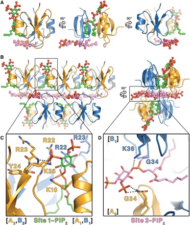
Fig. 2. Crystal structure of HBD-2 in complex with PIP2.

(A) The asymmetric unit comprises two HBD-2 molecules (cartoon representation; chain A in gold and chain B in blue) and two PIP2 molecules (ball and stick representations; oxygen in red, phosphorus in orange, and carbon in green for site 1 and pink for site 2). (B) Crystal packing of HBD-2 represented as in (A) as seen from the front (left) and from the side (right). (C) Close-up of site 1 represented as above, with PIP2 and all interacting residues shown as sticks. (D) Close-up of site 2 represented as above, with PIP2 and all interacting residues shown as sticks.
