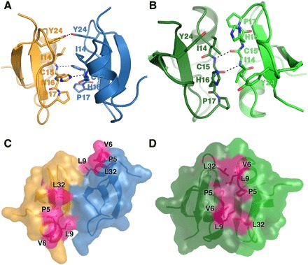
Fig. 4. Dimers of HBD-2 in the PIP2 bound and unbound state.

The dimer interfaces of HBD-2 are shown as cartoons, with residues 14 to 17 and 24 depicted as sticks and hydrogen bonds as dashed lines. (A) The PIP2 -bound HBD-2 dimer with chain A in gold and chain B in blue (PIP2 molecules are omitted for clarity). Two hydrogen bonds between H16 and the backbone of C15 are observed, as well as between the hydroxyl groups of Y24. (B) Apo–HBD-2 dimer with its two chains colored light green and dark green. Two hydrogen bonds between the backbones of C15 are shown. (C) Surface representation of (A). Exposed hydrophobic residues are shaded in pink. (D) Surface representation of (B). Exposed hydrophobic residues are shaded in pink.
