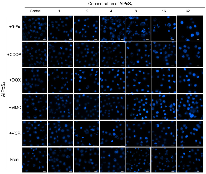
Figure 4.
Apoptosis induced by AlPcS4 + 5-FU, AlPcS4 + CDDP, AlPcS4 + DOX, AlPcS4 + MMC, AlPcS4 + VCR, and free-AlPcS4 in SGC-7901 cells after being irradiated for 12 h. The cells were treated with 1–32 µm/ml free-AlPcS4 or AlPcS4 + 5-FU (20 µm), AlPcS4 + CDDP (5 µm), AlPcS4 + DOX (0.4 µm/ml), AlPcS4 + MMC (0.5 µm/ml) or AlPcS4 + VCR (0.1 µm/ml) for 6 h. The cells were then irradiated with 635-nm laser irradiation at 100 mw/cm2 illumination dosage for 5 min, incubated for 12 h, stained with Hoechst 33342 probe, and then imaged using afluorescence microscope. All the Hoechst staining images were acquired at an ×400 magnification. The scale bar represented 20 µm. AlPcS4, Al(III) phthalocyanine chloride tetrasulfonic acid; 5-FU, 5-fluorouracil; DOX, doxorubicin; CDDP, cisplatin; MMC, mitomycin C; VCR, vincristine.

