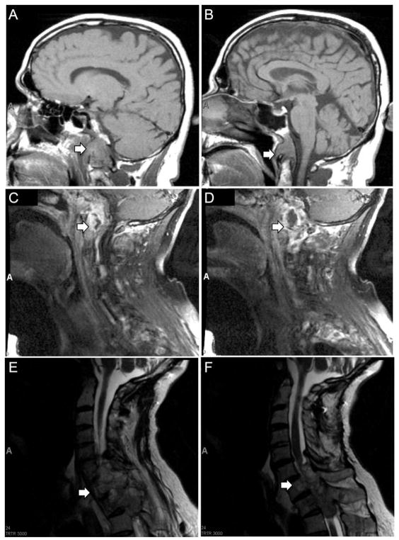
Figure 1. Magnetic resonance imaging of primary clival chordoma and recurrent tumor involving the cervical and thoracic spine.
(A,B) T1 sagittal MRI of brain demonstrating clival mass extending to inferior portion of odontoid (arrows), January 2007. (C,D) T1 sagittal cervical spine MRI demonstrating destructive lesion in soft tissues of skull base and upper cervical spine (arrows), May 2007. (E,F) T2 sagittal cervical spine MRI demonstrating extension of tumor from C5 to T2 with severe cord compression (arrows), September 2011.

