
Figure 5.
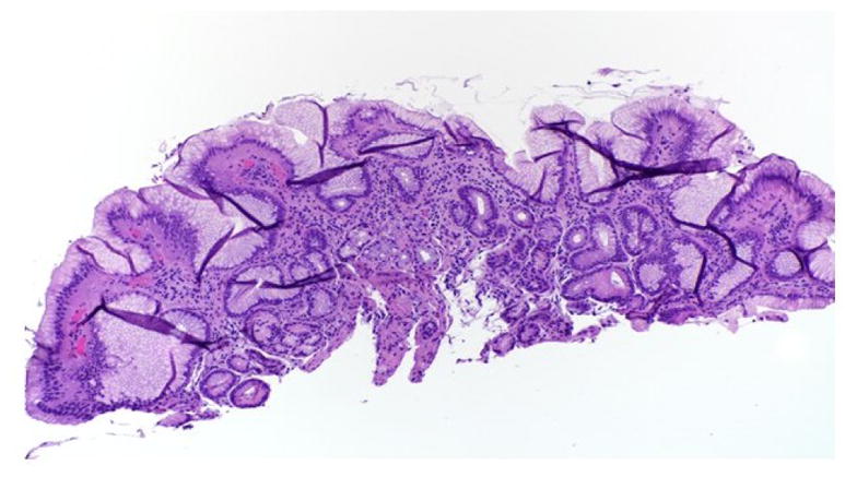
Low-power view shows columnar-lined epithelium, gastric pits, and underlying mucous glands with scattered chronic inflammatory cells within the underlying lamina classically seen with Barrett’s changes.
Official websites use .gov
A
.gov website belongs to an official
government organization in the United States.
Secure .gov websites use HTTPS
A lock (
) or https:// means you've safely
connected to the .gov website. Share sensitive
information only on official, secure websites.
Low-power view shows columnar-lined epithelium, gastric pits, and underlying mucous glands with scattered chronic inflammatory cells within the underlying lamina classically seen with Barrett’s changes.