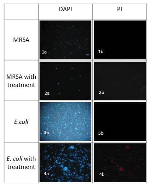
Figure 3.
Fluorescence Microscopy of bacterial cells treated with C16-K-RBB2 for 2h (Scale bar 10μm). (1a) Control, DAPI stained, no treatment with compound. (1b) Control, PI stained, no treatment with compound. (2a) MRSA, DAPI stained, treated with compound. (2b) MRSA, PI stained, treated with compound. (3a) Control, DAPI stained, no treatment with compound. (3b) Control, PI stained, no treatment with compound. (4a) E. coli, DAPI stained, treated with compound. (4b) E. coli, PI stained, treated with compound.

