Image 1.
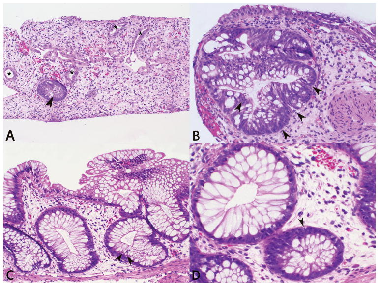
Photomicrographs of sigmoid colon biopsy with marked crypt drop out, lamina propria edema, crypts with damage/reactive epithelial changes (asterisks), and moderate epithelial apoptosis (arrows) (A, H&E, 10X; B, H&E, 20X). Photomicrographs of follow up colon biopsy with mild lamina propria edema, reparative changes, and rare epithelial apoptosis (arrows) (C, H&E, 20X; D, H&E, 40X).

