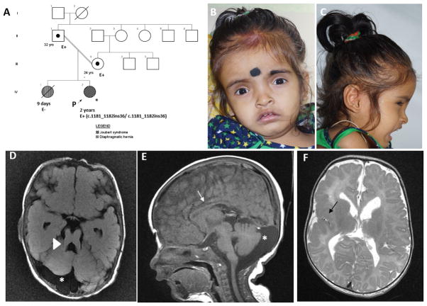
Fig 1.
Pedigree (A). Proband at age 2 years has frontal prominence, deep set eyes and midface hypoplasia (B, C). Magnetic resonance imaging of brain revealed thickening and lengthening of superior cerebellar peduncle (arrow head shows molar tooth sign, D), prominent cerebrospinal fluid space (asterix, D, E), thinning of corpus callosum (arrow, E), and perisylvian polymicrogyria (black arrow, F).

