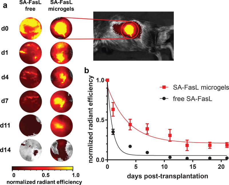
Figure 2. Microgels prolong SA-FasL retention in vivo.
SA-FasL was labelled with a near-IR dye and implanted under the kidney capsule of mice and imaged in vivo. (a) Representative images show localization of SA-FasL to graft site when presented on microgels, in contrast to diffuse signal measured in animals receiving free SA-FasL. Heat maps are consistent across animals in the same treatment group. Images are not shown for days 18 and 21 because signal was negligible. (b) Quantification of in vivo fluorescence and exponential decay curve fit demonstrate that microgels presenting SA-FasL prolong protein retention compared to free SA-FasL. Representative experiment (n=8 mice/group, mean ± SE; F-test (DFn, DFd) = 20.39 (2, 124) for curve fit parameters, two-tailed p < 0.0001) shown from 2 independent runs with consistent results.

