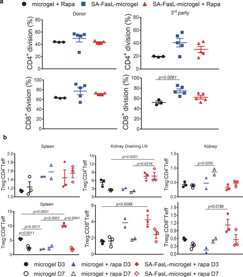
Figure 4. Immune monitoring and the role of CD4+CD25+FoxP3+ Treg cells in islet graft acceptance.
(a) Systemic response of long-term graft survivors to donor antigens. Carboxyfluorescein succinimidyl ester (CFSE)-labeled splenocytes were used as responders to irradiated BALB/c donor and C3H third party stimulators in an ex vivo mixed lymphocyte reaction assay. The dilution of CFSE dye in CD4+ and CD8+ T cells was assessed using antibodies to CD4 and CD8 molecules in flow cytometry and plotted as percent division for each cell population (n=3 mice/group for microgel and n=5 mice/group for microgel + rapa and SA-FasL-microgel + rapa; mean ± SE; ANOVA with two-tailed pair-wise comparisons using Tukey’s test). (b) Analysis of Teff and Treg cells. Single cells prepared from the spleen, kidney, and kidney-draining lymph nodes of the indicated groups on day 3 and 7 post-islet transplantation were stained with fluorescence-labelled antibodies to cell surface molecules that define CD4+ Teff (CD4+CD44hiCD62Llo), CD8+ Teff (CD8+CD44hiCD62Llo), and Treg (CD4+CD25+FoxP3+) populations and analyzed using flow cytometry. The ratios of Treg to CD4+ Teff and CD8+ Teff are plotted (n=2 mice/group for microgel + rapa and n=3 mice/group for microgel and SA-FasL-microgel + rapa; mean ± SE; ANOVA with two-tailed pair-wise comparisons with Bonferroni’s correction).

