Figure 4.
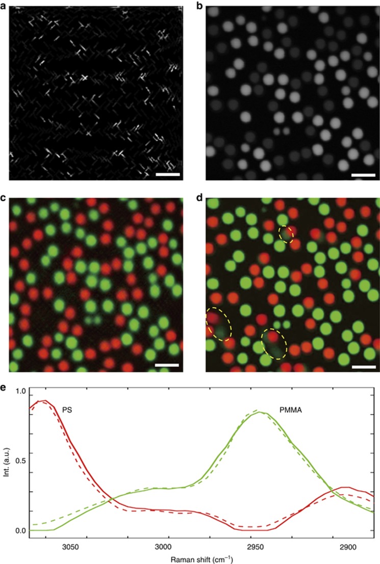
Experimental results for PS and PMMA microbead mixture in water. (a) One frame of the sparsely sampled raw spectroscopic image at 2915 cm−1 with a pixel dwell time of 2 μs; the entire spectroscopic SRS data cube with 50 frames was captured in 0.8 s. (b) One frame of the raster-scanned spectroscopic SRS image at 2915 cm−1; the stack was captured at a speed of 2 frames s−1. Output concentration maps using regularized spectroscopic image unmixing for the (c) sparsely sampled image and (d) raster-scanned image, respectively. Motion artifacts induced by high bead motility are shown in d (selected examples are highlighted by yellow circles). (e) Output spectral signatures for sparsely sampled image (solid line) and raster-scanned image (dotted line). Scale bars, 10 μm.

