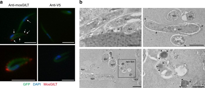
Fig. 3.
MosGILT binds to the surface of P. berghei sporozoites. a Immunostaining with a mosGILT specific monoclonal antibody revealed endogenous SG mosGILT bound to the surface of P. berghei sporozoites. An irrelevant mouse monoclonal, anti-V5, was used as a control. All images are representative of more than three independent experiments. MosGILT bound to sporozoites is highlighted by white arrows (Scale - 5 μm). b Immunoelectron microscopy labeling of endogenous mosGILT on P. berghei sporozoites within intact SGs. Sporozoites labeled with a control mouse serum (top left). Sporozoites labeled with the anti-mosGILT monoclonal antibody, top right and bottom panels. The region highlighted by a black square represents a higher magnification of predicted sporozoite anterior or posterior tips, bottom right. Arrows highlight mosGILT labeling at the surface of sporozoites (spz-sporozoite; scale 1 μm and 500 nm bottom right)

