Figure 3.

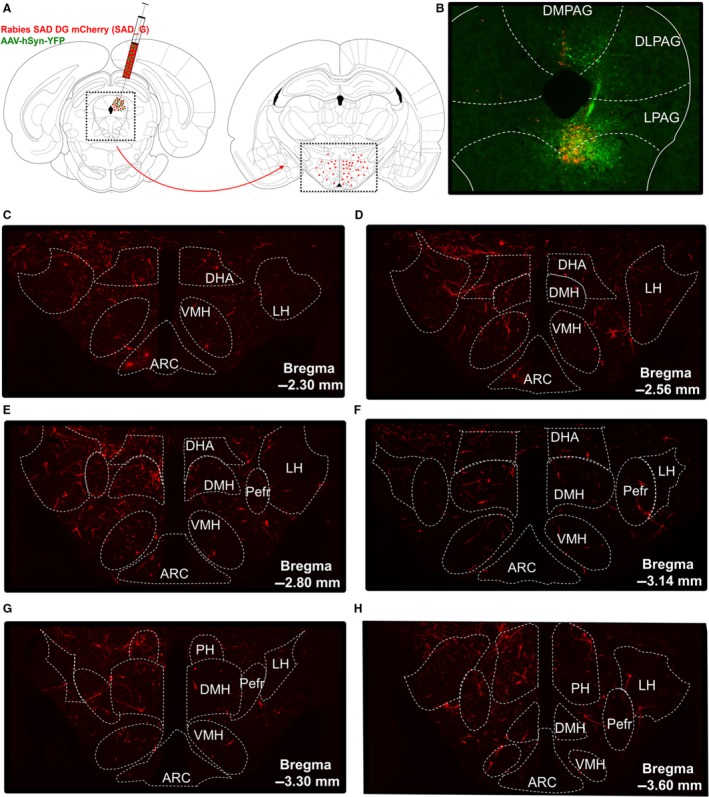
Retrograde tracing from PAG to hypothalamus. (A) Experimental design. Rabies SAD DG mCherry (SAD_G), a monosynaptic retrograde tracer virus, was unilaterally injected into the PAG together with an AAV‐hSyn‐YFP virus to visualize the injection site. The number of direct presynaptic inputs in defined subregions of the hypothalamus, including the DMH, was assessed. (B) Representative injection site, showing the needle track and most GFP‐positive cell bodies (green) in the ventral PAG. (C–H) Direct presynaptic inputs (red) in the anterior‐to‐posterior hypothalamus for the representative injection site in B. Distances from bregma (mm) are indicated, and the dotted outline shows the boundaries of hypothalamic subregions in which the number of presynaptic inputs was counted. ARC, arcuate nucleus; DHA, dorsohypothalamic area; DMH, dorsomedial hypothalamus, LH, lateral hypothalamus; Pefre, perifornical area; PH, posterior hypothalamus; VMH, ventromedial hypothalamus.
