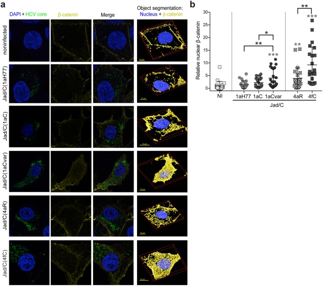
Figure 6.
Strain-specific, core-dependent nuclear translocation of β-catenin in infected cells. (a) Huh-7.5 cells infected with the indicated viruses or noninfected cells were labeled for nucleus (DAPI, blue) and core (green), as well as for β-catenin (yellow). Merged deconvolved images and representative 3D segments used for object segmentation (nuclei and β-catenin, right images) are shown. (b) Images were subjected to object analysis (co-localization intersection) using Huygens Professional software and intersecting volumes between β-catenin and nucleus were quantified per cell. Intersecting voxels (means and distribution among 23 cells per condition, each cell being represented by a symbol) were expressed relatively to intersecting voxels found in noninfected cells set at 1. Statistical analyses with respect to values obtained in noninfected cells are indicated above each group of virus-infected cells (in grey characters), while statistical analyses between two related variants are indicated in black characters above brackets. These statistical analyses were performed according to the Holm-Sidak method and are coded as follows: P < 0.05 (*), P < 0.005 (**), P < 0.001 (***).

