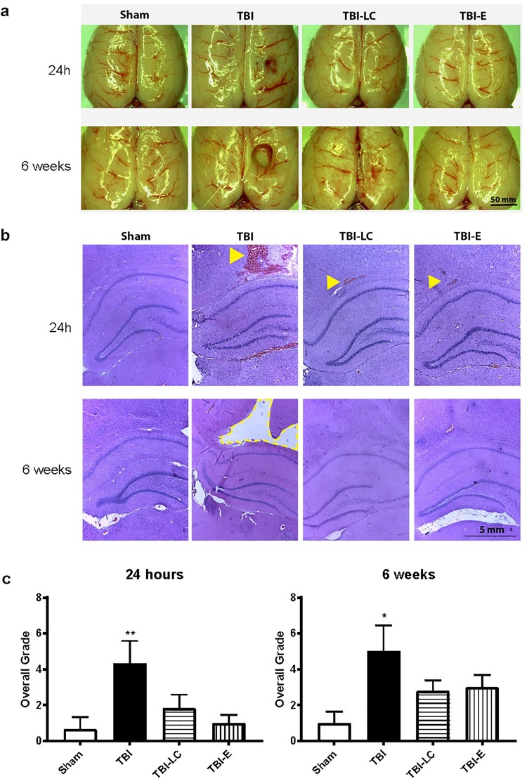
Figure 2.
Dorsal images of brain (a), representative haematoxylin and eosin-stained coronal sections of the right brain at lesion level (b), and semi-quantification of injury in the coronal brain sections by overall grade (c) at 24 hours and 6 weeks post-surgery. In (b), signs of injury included tissue disruption, haemorrhage (arrowheads) and tissue loss (dotted line) presented as a cavity (TBI at 6 week). Data was analysed by One-way ANOVA with Bonferroni post hoc tests, n = 6, *P < 0.05, **P < 0.01 vs Sham. TBI: Traumatic brain injury; TBI-LC: Traumatic brain injury with L-Carnitine treatment; TBI-E: Traumatic brain injury with Exendin-4 treatment.

