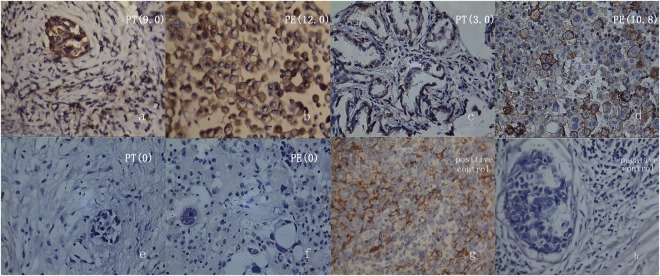
Figure 1.
PD-L1 expression in pleural tissue (PT), pleural effusions (PE) of pulmonary adenocarcinoma by immuhistochemical stain (400 times magnification). The slices of (a–f) are matched respectively by PT and PE from the same patients. If 5 points is the cut-off value, PD-L1 expression of PT and PE are both positive in the first case, a negative and a positive in the second case, both negative in the third case. The (g and h) are positive (lymphoma) and negative (breast carcinoma) control. The values in brackets are the mean scores in each slice of five scopes.

