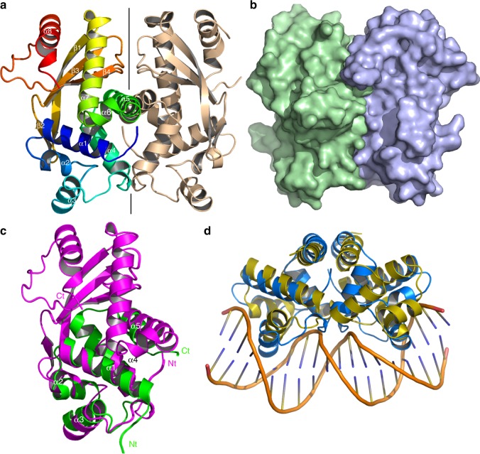
Fig. 3.
Crystal structure of AcrIIA6. a Ribbon view of the AcrIIA6 dimer. The left monomer is rainbow-colored, the right monomer is colored in beige. The secondary structures are identified α1–α8, β1–β4. b Surface representation with the same orientation and colors as in a. c Superimposition of AcrIIa6 monomer A (magenta) on a monomer of a putative transcription factor (PDB 2ef8, green). Helices α1–α5 are structurally aligned. d Superimposition of the AcrIIa6 dimer (blue) on the controller protein C.Esp1396I operator (yellow) in complex with dsDNA (orange; PDB 3s8q37). Both helices α3 are inserted in adjacent major grooves

