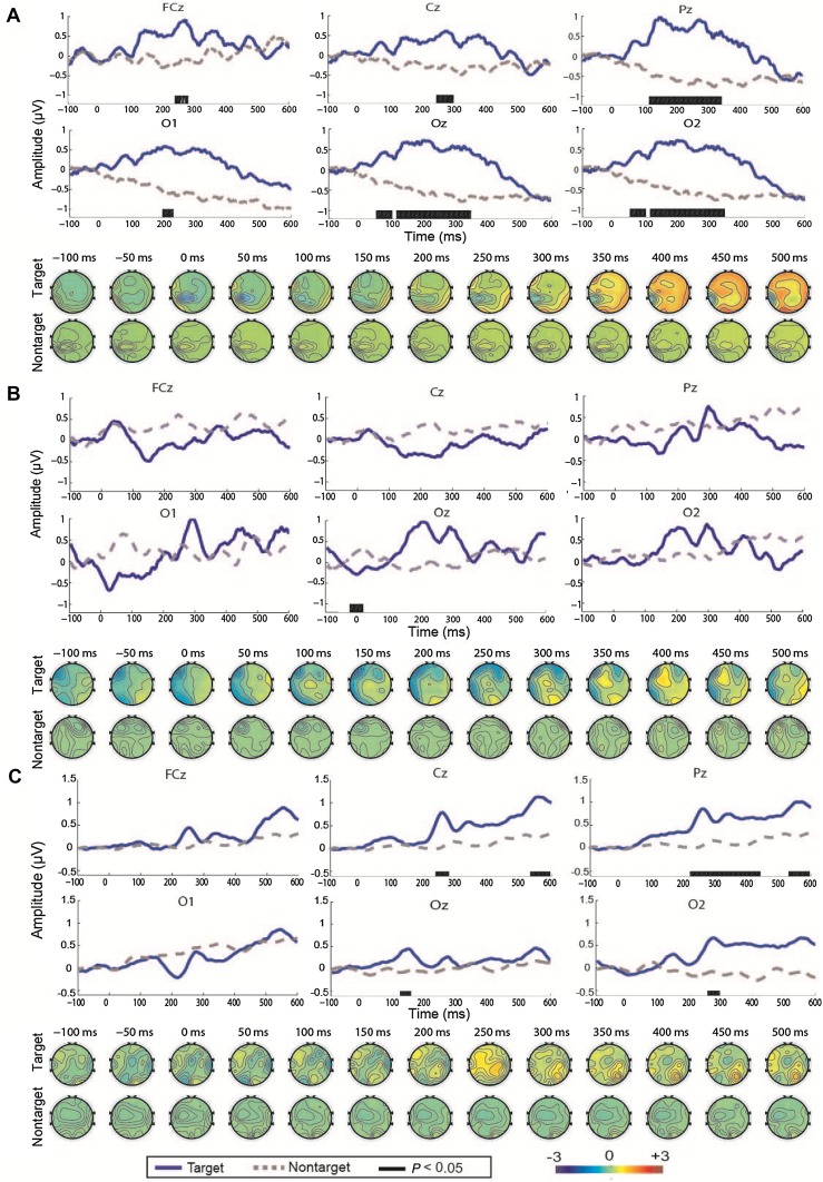
Fig. 3.
A–C ERP waveforms and scalp topography in patients 12 (A), 13 (B), and 15 (C). The waveforms were obtained from 100 ms before the stimulus onset to 600 ms after the stimulus onset in six channels (FCz, Cz, Pz, O1, Oz, and O2). The blue solid and gray dashed lines denote the waveforms evoked by the target and non-target stimuli, respectively.

