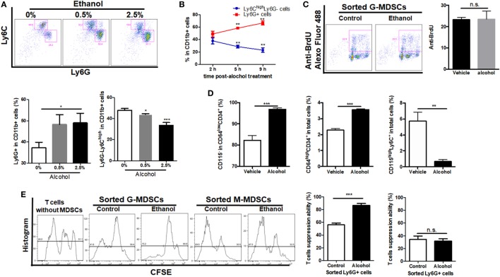
Figure 4.
Effect of ethanol on the differentiation, expansion, and function of myeloid-derived suppressor cells (MDSCs) populations in vitro. Bone marrow-derived cells were isolated from naïve mice and treated directly with 0.5 or 2.5% ethanol in RPMI-1640 medium supplemented with 10% FBS. After culturing for 2, 5, and 9 h, cells were collected for the examination of MDSCs population via flow cytometer. (A) Representative images and quantification of flow cytometric analyses granulocytic-MDSCs (G-MDSCs) populations treated with ethanol at different concentrations. (B) Time course of population of G-MDSCs and monocytic-MDSCs (M-MDSCs) cultured with ethanol at the concentration of 0.5% (vol/vol). (C) Representative images and quantification of flow cytometric analyses anti-BrdU Alexo Flour 488 staining. Bone marrow-derived cells were labeled with BrdU at a final concentration of 10 µM. Then, cells were analyzed by flow cytometer. The proliferation of G-MDSCs was not increased by ethanol treatment. (D) Populations of common myeloid precursor, granulocyte-monocyte progenitors (GMPs), and monocyte-committed progenitors produced by GMPs (MP) in bone marrow-derived cells treated with ethanol or vehicle determined by flow cytometer. (E) Representative images of flow cytometric analyses of carboxyfluorescein succinimidyl ester (CFSE) intensity in CFSE-CD8+ T cells co-cultured with G-MDSCs populations or M-MDSCs treated with ethanol or vehicle. Data were analyzed as mean value ± SD and Student’s T-test was used to assess the result significance. *p < 0.05, **p < 0.01, ***p < 0.001, compared with the control group; n.s., not significant.

