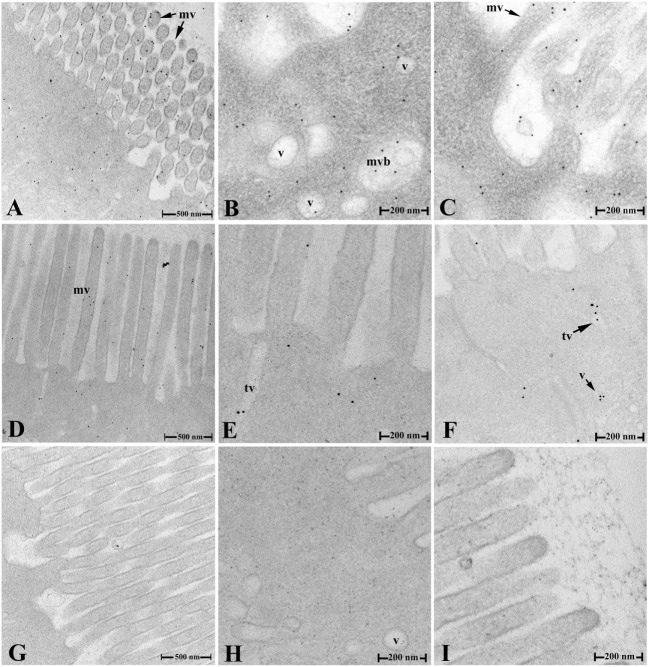
Figure 4.
Transepithelial transport of the OVA evidenced by IEM. (A–C) The distribution of the OVA-positive gold particles in various sized vesicles, tubules, and multivesicular bodies in the cytoplasm, around the surface microvilli of IECs and in gut lumen in group 1 at 2 h after challenge. (D–F) The distribution of the OVA-positive gold particles in IEC of group 1 at 9 h after challenge. (G–I) No gold particles were detected in negative control staining (G) and IECs of hindgut in group 2 (H,I) at 2 h after challenge. Abbreviations: v, vesicles; tv, tubule vesicles; mvb, multivesicular bodies; mv, microvilli; IECs, intestinal epithelial cells; OVA, ovalbumin; IME, immunogold electron microscopy.

