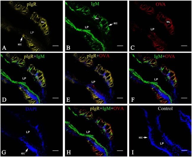
Figure 5.
Co-localization of pIgR, IgM, and OVA in hindgut of flounder. Pictures from group 1 were shown as examples. The fish immunized intraperitoneally with OVA and subsequently challenged with OVA via caudal vein on the fourth week after immunization, and pIgR, IgM, and OVA in hindgut was stained at 2 h after challenge, presenting orange, green and red fluoresce respectively. The cell nucleus was counterstained in blue with DAPI. (A,B,C) Distribution of IgM, OVA, pIgR in hindgut at 2 h after challenge was illuminated, respectively. (D–F) The merged picture of (A,B,G) and (A,C,G) and (B,C,G), respectively. (G) The cell nucleus of intestinal epithelium was stained blue by DAPI. (H) The merged picture of (A–C,G); (I) Negative control. Bar = 25 μm. Abbreviations: LP, lamina propria submucosa; Lu, lumen; IECs, intestinal epithelial cells; Mu, mucus cells; BV, blood vessel; OVA, ovalbumin.

