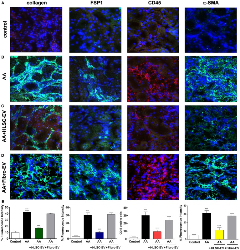
Figure 3.
Immunofluorescent staining of kidneys from mice treated with aristolochic acid (AA). Kidney cryo-sections from healthy mice (A), mice treated with AA (B), AA mice treated with HLSC-EVs (C), or Fibro-EVs (D) were stained for collagen 1a1, fibroblast-specific protein 1 (FSP-1), CD45, and α-SMA to identify presence of tissue fibrosis, fibroblasts, and inflammatory cells (Original magnification: 200×). (E) Histograms depicts the fluorescence intensity of collagen, FSP-1, α-SMA, and quantification of cells positive for CD45 in mouse kidney cryo-sections from AAN mice experimental groups. Data represent mean ± SD of the fluorescence intensity or cells positive per high power field measured from 10 images taken at random from six samples per treatment (n = 6 mice). ***p < 0.001 AA vs control or HLSC-EVs vs AA. No significant differences were observed between AA vs Fibro-EVs.

