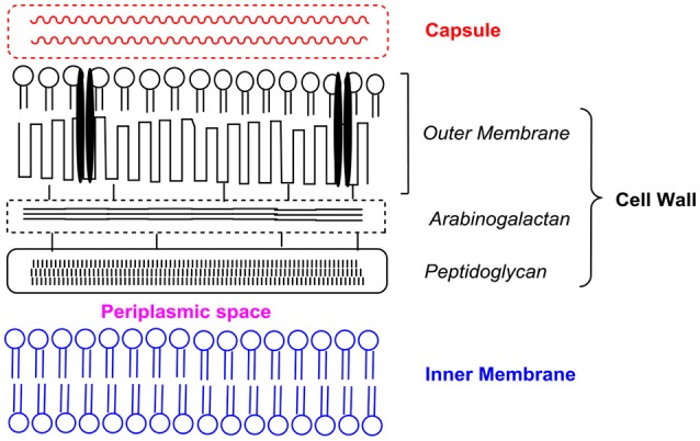
Figure 2.

Schematic representation of the different components of the mycobacterial cell envelope based on the description given by Daffé et al. (2017). Presentation is not drawn to scale. Interspersed within the outer membrane are porin channels (depicted as black parallel bars) for the uptake of small hydrophilic molecules.
