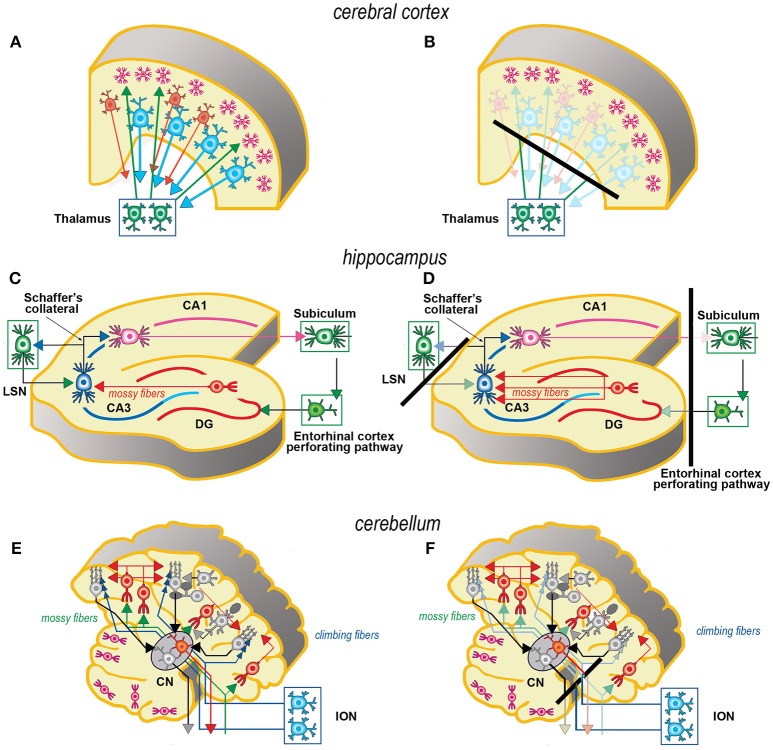
Figure 1.
Simplified schematic representations of the effects of the slicing procedure on the preparation of acute slices (left) and OCs (right) from cerebral cortex (A, B), hippocampus (C, D), and cerebellum (E,F). Only the main components of circuitry are depicted as well as the principal afferent and efferent connections. The lines of section (solid black bars) of the main fiber systems are represented only in the right panels to show the effects of resection onto the afferent and efferent systems (shadowed in comparison to the corresponding drawings at left). The magenta neurons in E, F represent the external granular layer of the cerebellar cortex, a temporary subpial cell layer that disappears in the mature cerebellum. CN, cerebellar nucleus; CA1, CA3, hippocampal subfields CA1 and CA3 (from Cornu Ammonis); DG, dentate gyrus; ION, inferior olivary nucleus; LSN, lateral septal nucleus.

