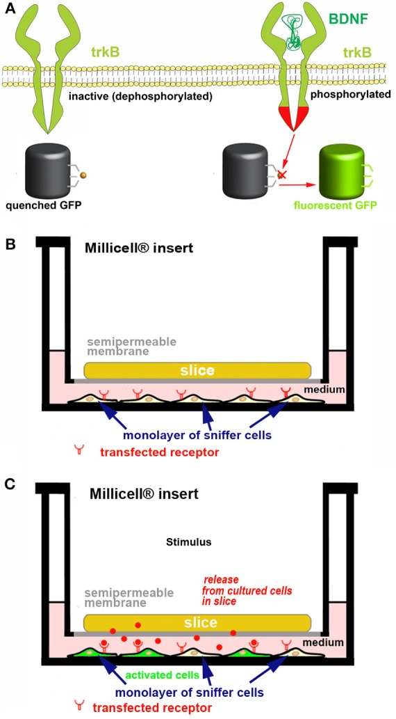
Figure 6.

Strategy for developing a new ex vivo platform to study the effect of a neurotropic factor. The example describes a co-culture made of an acute or organotypically cultured brain slice and a monolayer of sniffer cells seeded at the bottom of a Petri dish. If an acute slice is used it needs to be kept in place with an ad hoc grid (not represented for simplicity). (A) engineering of the sniffer cell line to make it suitable for reporting the release of BDNF from slice cells. The sniffer cells are double transfected to express trkB receptors and a dark-to-bright state GFP-based biosensor (60, 61) that is modified to become activated after trkB phosphorylation. (B,C) Scheme of the co-culture set-up. In (B) the sniffer cells are not fluorescent, whereas in (C) the BDNF released from the slice (red dots) diffuses through the culture medium, binds to trkB receptors on the sniffer cells and thus some of these cells become fluorescent. The process can be imagined in real time using an inverted wide field or confocal microscope. In the example it is assumed that BDNF release is triggered after an ad hoc stimulus.
