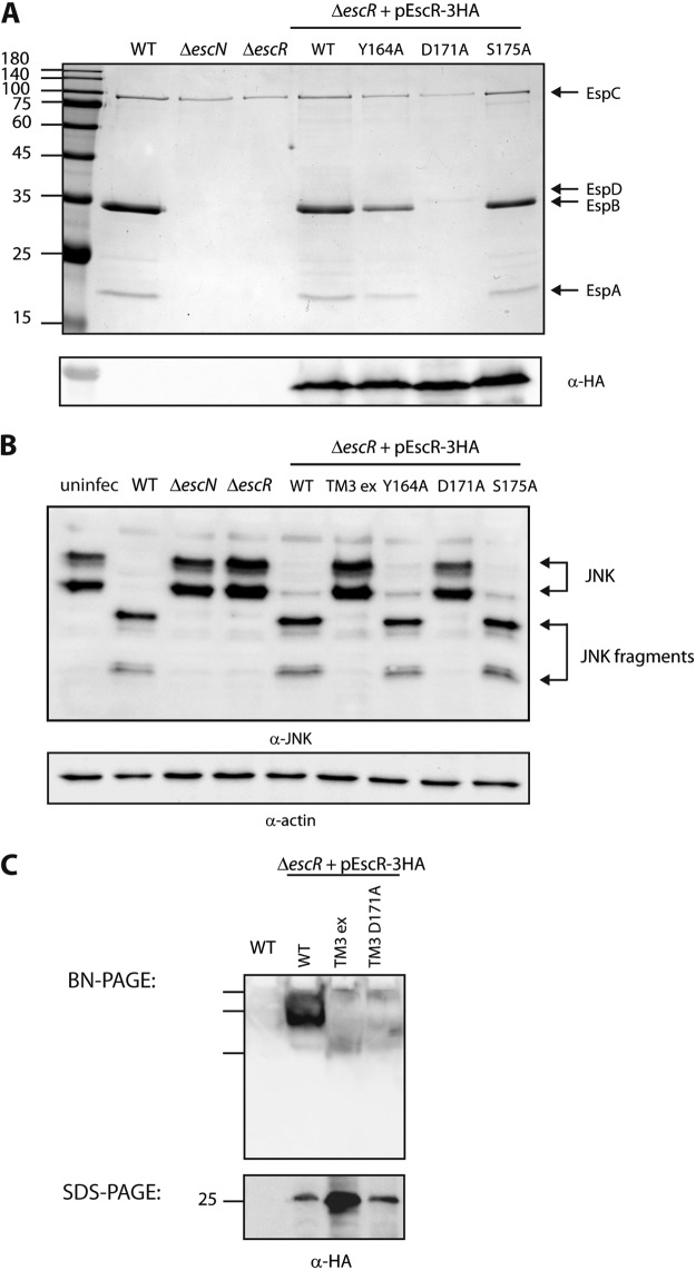
FIG 6 .
The aspartic acid residue found in the predicted EscR TMD3 sequence is critical for T3SS activity and the ability of the bacteria to infect host cells. (A) Protein secretion profiles of EPEC strains grown under T3SS-inducing conditions: WT, ΔescN and ΔescR strains, and ΔescR strain complemented with EscRWT-3HA, EscRY164A-3HA, EscRD171A-3HA, or EscRS175A-3HA. The secreted fractions were treated and analyzed as described in the Fig. 1A legend. A point mutation at position 171 of EscR TMD3 (D to A) abolished T3SS activity, while point mutations at position 164 or 175 (Y to A or S to A, respectively) demonstrated active T3SS. The expression of EscR-3HA variants was examined by analyzing the bacterial pellets by SDS-PAGE and Western blot analysis with an anti-HA antibody. Numbers at left are molecular masses in kilodaltons. (B) HeLa cells were infected with one of the following EPEC strains: WT, ΔescN or ΔescR strain, or ΔescR strain complemented with EscRWT-3HA, EscR-TM3ex-3HA, EscRY164A-3HA, EscRD171A-3HA, or EscRS175A-3HA. JNK and its degradation fragments are indicated at the right of the gel. WT EPEC showed massive degradation of JNK similarly to the ΔescR strain complemented with EscRWT-3HA, EscRY164A-3HA, or EscRS175A-3HA. However, ΔescR EPEC strains transformed with EscR-TM3ex-3HA or EscRD171A-3HA showed the same JNK pattern as the uninfected sample and the samples infected with ΔescN or ΔescR mutant strains. (C) Membrane protein extracts of WT EPEC and ΔescR mutant complemented with EscRWT-3HA, EscR-TM3ex-3HA, or EscRD171A-3HA were incubated in BN sample buffer and then subjected to BN-PAGE (upper panel) and SDS-PAGE (lower panel) and Western blot analysis using anti-HA antibody. (Upper panel) BN-PAGE analysis showed that EscRWT-3HA forms a high-molecular-weight protein complex that is absent from the EscR-TM3ex-3HA and the EscRD171A-3HA samples. We observed only three marker bands in the Western blot of BN-PAGE; therefore, we cannot estimate the size of the complex. (Lower panel) To confirm similar EscR expression levels among the samples, membrane protein extracts were analyzed by SDS-PAGE and Western blot analysis using anti-HA antibody. Similar protein expression levels were observed.

