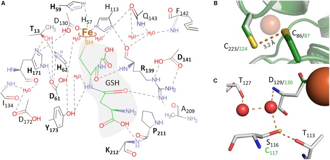
FIGURE 5.
Molecular representations of the AcPDO 3D model with glutathione from the PpPDO structure (5VE5). (A) Theoretical model of GSH in the AcPDO active site structure and predicted hydrogen bonds; boldface amino acid residues were mutagenized in this study. (B) C86 and C223 in the MxPDO 3D structure (gray) and in the AcPDO model (green). (C) Comparison of the secondary coordination sphere around D129/130 between the MxPDO (gray) and the AcPDO (green) originating from the S116 residue in the MxPDO.

