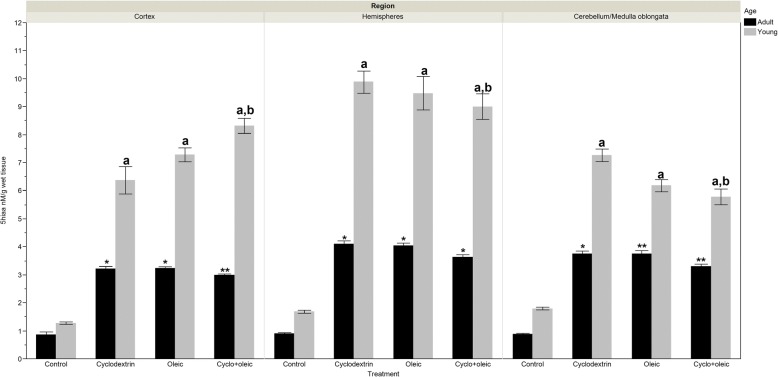
Fig. 1.
Levels of 5-hydroxyindol acetic acid (5-HIAA) in brain regions of adult and young Wistar rats treated with oleic acid and cyclodextrin. Adult rat. Cortex: Anova F = 240.29 p < 0.0001. *) p < 0.0001 vs control, **) p = 0.047 vs Oleic. Hemispheres: Kruskal-Wallis X2 = 41.57 p < 0.0001. *) p < 0.0001 vs control, **) p = 0.026 vs Cyclodextrin. Cerebellum/ Medulla Oblongata: Kruskal-Wallis X2 = 43.39 p < 0.0001. a) p < 0.0001 vs control, **) p = 0.013 vs Oleic and p = 0.009 vs Cyclodextrin
Young rat. Cortex: Kruskal-Wallis X2 = 47.54 p < 0.0001. a) p < 0.0001 vs control, a,b) p = 0.039 vs Oleic, and p = 0.017 vs Cyclodextrin. Hemispheres: Kruskal-Wallis X2 = 42.07 p < 0.0001. a) p < 0.0001 vs control. Cerebellum/medulla oblongata: Kruskal-Wallis X2 = 49.75 p < 0.0001. a) p < 0.0001 vs control, a,b) p < 0.04 vs Cyclodextrin.

