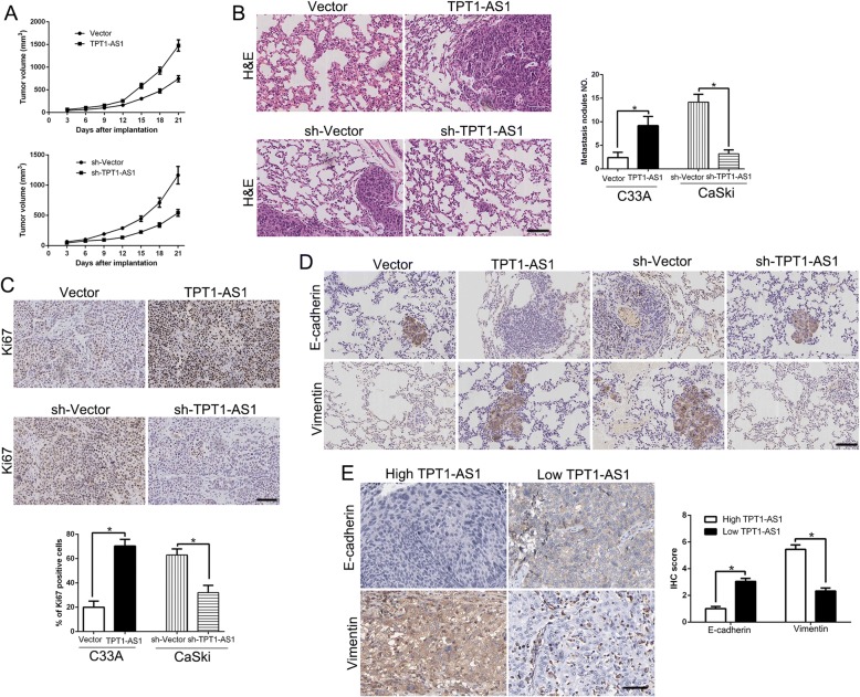
Fig. 3.
TPT1-AS1 promotes tumor growth and metastasis in vivo. a Tumor growth curve revealed that TPT1-AS1 overexpression significantly promoted, while TPT1-AS1 knockdown inhibited tumor growth in vivo (n = 5). b Representative HE staining of lung metastases in TPT1-AS1 overexpression or knockdown cells (n = 5). c Tumor nodules were subjected to immunohistochemical staining for Ki-67 assays and quantitative analysis. Representative immunostaining assays revealed that TPT1-AS1 overexpression significantly increased the number of Ki-67 positive cells. However, the percentage of Ki-67 positive cells in tumors arising from the TPT1-AS1 knockdown group was significantly lower than that in the negative control group. d Immunohistochemistry of E-cadherin and Vimentin were showed and compared between tissues of respective TPT1-AS1 expression level. e Immunohistochemistry of E-cadherin and Vimentin were showed and compared between TPT1-AS1 high expressing CC tissues (n = 59) and TPT1-AS1 low expressing cases (n = 56). *P < 0.05

