
Figure 4.
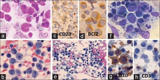
Centroblastic lymphoma: A 37-year-old man presented with 2 × 2 cm submental lymph node and 1 × 0.5 cm pre-auricular lymph node. Clinical diagnosis: lymphoma versus nasopharyngeal carcinoma. Cytodx: NHL: centroblastic lymphoma/large B-cell lymphoma, ICC: CD20+, BCl2+, histopathological diagnosis: NHL: follicular Gr II. (a) Large lymphoma cells with cleaved- or noncleaved nuclei (MGG × 1000). (b) Lymphoma cells with prominent central or peripheral nucleoli (Pap × 1000). (c) Lymphoma cells with cytoplasmic positivity for CD20 (×400). (d) Lymphoma cells with cytoplasmic positivity for BCl2 (×1000). (e-h) Diffuse large B-cell lymphoma (DLBCL): A 60-year-old man presented with 3 × 2 cm right axillary lymph node of >2 months duration. FNA cytodiagnosis: TCRBCL, ICC: CD20+ (tumor cells), CD3+ (lymphocytes), CD30–, and EMA–. Histopathological diagnosis: DLBCL. (e) Lymphoma cells mixed with numerous mature lymphocytes (Pap × 400). (f) Large lymphoma cells and lymphoglandular bodies (MGG × 1000). (g) Lymphoma cells positive for CD20 (×1000). (h) Mature lymphocytes positive for CD3 (×1000)