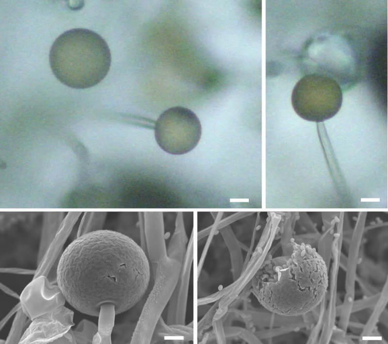
Figure 2. Sporangia containing numerous sporangiospores.

The SEM image shows intact sporangia (top panel). Each sporangium can produce several sporangiospores (bottom panel). Scale = 20 μm (adapted from Li et al., (2011) Sporangiospore size dimorphism is linked to virulence of Mucor circinelloides. PLOS pathogens).
