. 2016 Jul 13;5(5):1445–1452. doi: 10.1039/c6tx00216a

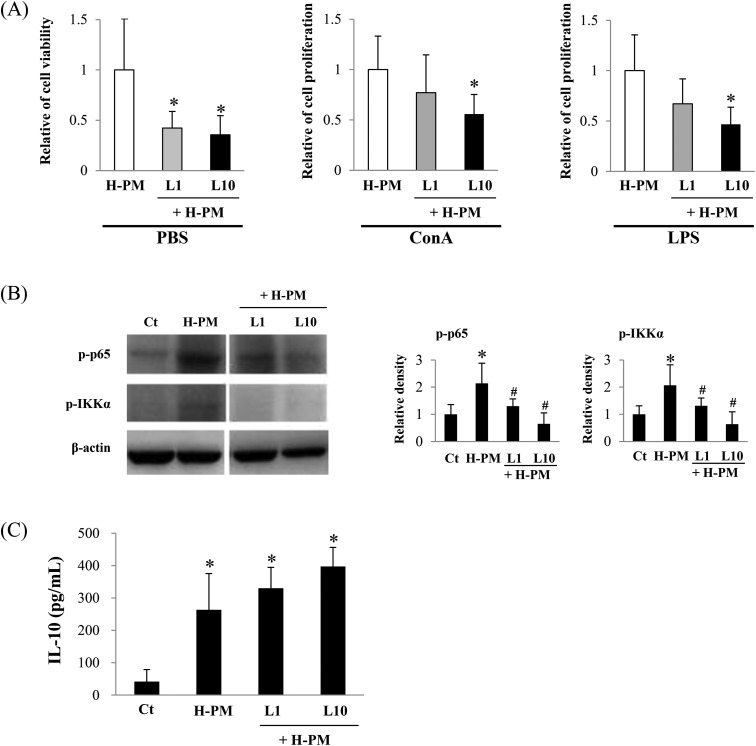
Fig. 4. LPS inhibits H-PM-mediated NF-κB activation in vivo. Saline (Ct) or H-PM in the presence or absence of LPS (1 ng per mouse [L1] or 10 ng per mouse [L10]) was intratracheally administered to BALB/c mice, and the mice were dissected at 24 h after the last administration. (A) Cell proliferation was measured after cell culture for 72 h as described in the legend of Fig. 1. *p < 0.05 vs. H-PM-administered mice (n = 7 in each group). The values shown are the mean relative cell proliferation as compared to that of the H-PM group. (B) Whole cell lysates of freshly isolated splenocytes were prepared and probed for the activation of NF-κB by performing western blot analysis. The levels of p-p65 and p-IKKα are shown. β-Actin was used as a loading control. In the right panel, quantification of the western blot data is shown. The values shown are the mean of the relative density ± SD after normalization to β-actin levels. Representative blots are shown. *p < 0.05 vs. control (n = 4 in each group). (C) Splenocytes (2 × 10⁵) were treated with LPS and supernatants were collected at 12 h after culture. IL-10 levels were measured by ELISA. *p < 0.05 vs. control (n = 5 in each group).

Fig. 4