. 2016 May 17;5(4):1172–1181. doi: 10.1039/c6tx00084c

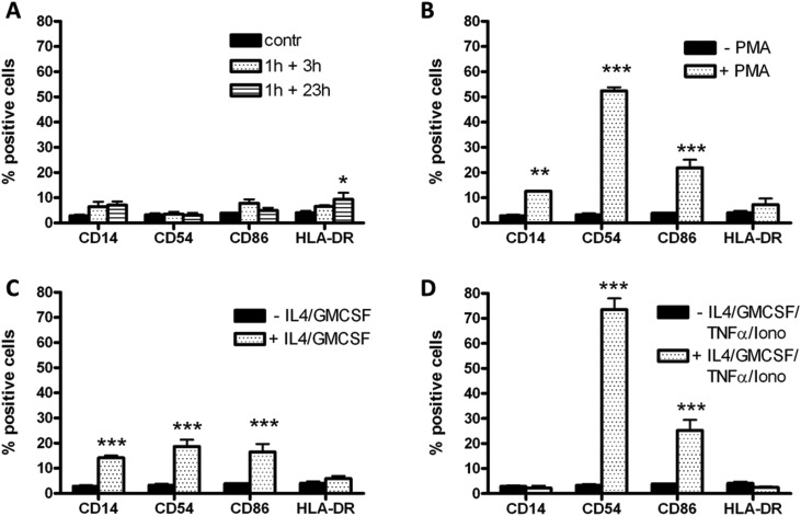
Fig. 5. Effects of PLTX-treated HaCaT conditioned media on monocyte differentiation. (A) Cells were exposed for 120 h to HaCaT conditioned media (1 h exposure to 1.0 × 10⁻¹¹ M PLTX followed by 3 and 23 h culture in toxin-free medium) and the levels of the following differentiation markers CD14, CD54, CD86 and HLA-DR evaluated by flow cytofluorometry. Panel B, C and D: positive controls. Monocytes were differentiated into (B) macrophages (10–7 M PMA), (C) iDCs (100 ng per mL IL-4 and GM-CSF) and (D) mDCs (200 ng per mL IL-4, 100 ng per mL GM-CSF, 10 ng per mL TNF-α and 200 ng per mL ionomycin). The results are expressed as % of positive cells to the markers considered and are the average of 4 experiments performed in duplicate (black bars: undifferentiated monocytes; dotted bars: differentiated cells). Statistical differences vs. undifferentiated monocytes: *, p < 0.05; **, p < 0.01; ***, p < 0.001 (one-way ANOVA and Bonferroni post-test).

Fig. 5