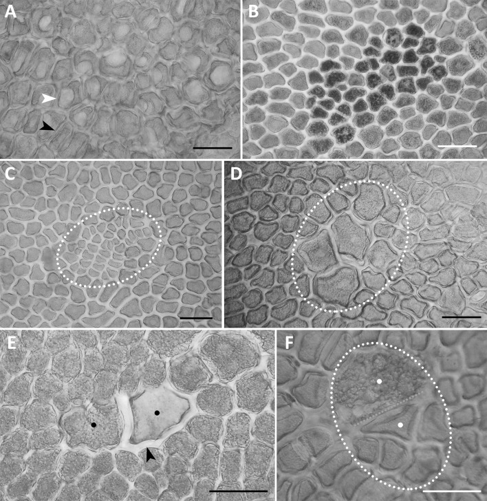
Fig. 4.
Examples of variability of an isolated aleurone layer (a surface view). a Vacuolar (white arrowhead) and a-vacuolar (black arrowhead) parts of the aleurone layer in A. sativa; b a group of dark aleurone cells with multiple globoids in the amphiploid A. abyssinica × A. strigosa; c a clone of small aleurone cells (outlined) with a short cell cycle in the amphiploid A. barbata × A. sativa ssp. nuda; d a clone of large aleurone cells (outlined) with a long cell cycle in the amphiploid A. fatua × A. sterilis; e two phenotypes (black dots) of polyploid sister aleurone cells after somatic crossing-over—the left cells with normal aleurone grains and cell wall, the right with a thick hemicellulosic wall (black arrow), and light aleurone grains in the amphiploid A. abyssinica × A. strigosa; f two sister cells (outlined and white dots) in the aleurone layer expressing two phenotypes; the lower cell is ‘aleurone’ and higher is ‘starchy’; after somatic crossing-over in the amphiploid A. barbata × A. sativa ssp. nuda. a–f documented in a polarising microscope at differently crossed nicols. Scale bars 100 µm

