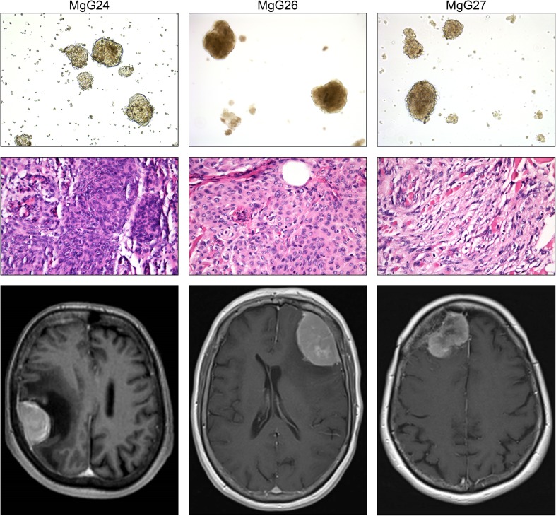
Fig. 2.
Top panel shows micrographs of 3D meningioma cultures with × 10 magnification. Middle panel depicts micrographs of H&E stained original patient tumour at × 40 magnification. Bottom panel are gadolinium-enhanced MRI scans. 3D cultures showed aggregated cell formation into a sphere. H&E stained tumour samples confirmed the diagnosis of meningioma in all three cases. MRI scans revealed meningiomas at the convexity and skull base

