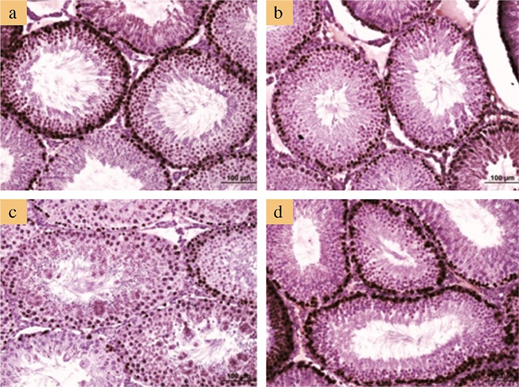
Figure 4. Representative micrographs of proliferating cell nuclear antigen immunostaining. Positive staining notably occurred in spermatogonium and spermatocytes in the control group (a); similar staining was observed in the L-cysteine group (b). Note that proliferating cell nuclear antigen immunostaining decreased notably in seminiferous tubules containing multinucleated giant cells in the acrylamide alone group (c), and it is restored in the acrylamide+L-cysteine group (d). Bars represent 100 μm.

