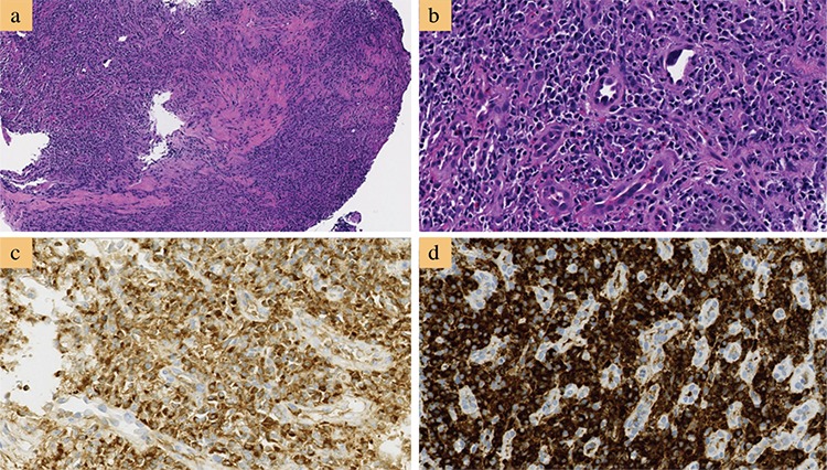
Figure 2. Histologic examination of one classic immunoglobulin G4-related esophagitis. Histologic section shows dense lymphoplasmacytic inflammation rich in plasma cells with storiform fibrosis and obliterative phlebitis (hematoxylin and eosin) stain, a) 40×; b) 400×. Majority of the plasma cells are positive for IgG, c) and immunoglobulin G4, d) (immunohistochemistry, 400×, each.). Reprinted from Obiorah et al. (64). Reprinted with permission of Oxford University Press.

