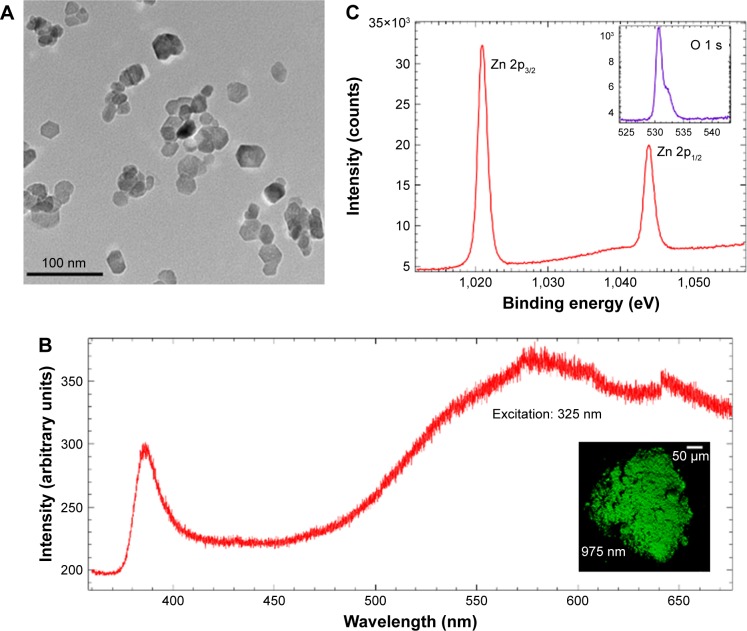
Figure 1.
(A) A representative transmission electron micrograph for ZnO NPs prepared using the polyol method. (B) Single-photon excitation ~325 nm (resonant with ZnO band gap) photoluminescence from ZnO NPs shows ultra-violet (UV) emission arising from the band-edge along with more intense broad visible emission from intrinsic defects between 500 and 650 nm. The inset shows a multiphoton microscope image of Zn NPs visible luminescence excited with ~975 nm three-photon equivalent of ZnO band gap. (C) X-ray photoelectron spectra of Zn and O (shown in the inset) showed that the as-prepared ZnO NPs are non-stoichiometric with Zn vacancies.

