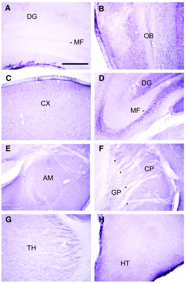
Fig. 3.
Sections from the forebrain. a Sections labelled with antigen-absorbed antibody, showed absence of labelling. b Moderately dense staining is present in the olfactory bulb. c The cerebral cortex (CX) is moderately labelled. Staining is present in punctuate profiles in the neuropil. d The cell bodies, dendrites (DG) and axons (mossy fibres, MF) of dentate granule neurons are densely labelled. e The amygdala (AM) is lightly labelled. f The caudate-putamen (CP) is lightly labelled. The globus pallidus (GP) is very lightly labelled or unlabelled. g The thalamus is lightly labelled. h The hypothalamus (HT) is moderately densely labelled. Scale = 0.5 mm

