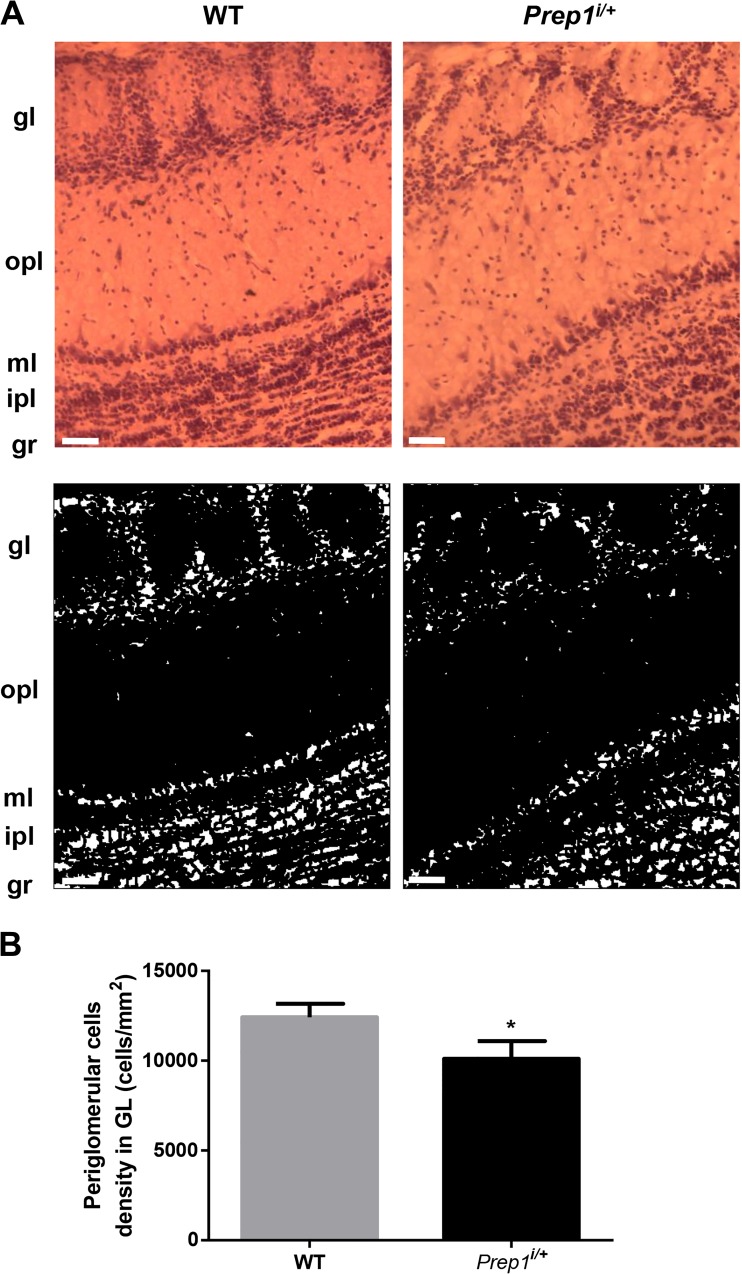
Fig. 4.
Hemalum staining of Prep1i/+ mice OB sections. a Representative images of olfactory bulb coronal cryosections from WT and Prep1i/+ mice stained with Hemalum staining, a solution of hematoxylin and alum able to stain cell nuclei (scale bar, 50 μm). A threshold was applied to the images in order to identify and count the nuclei (lower panel). b Quantification of neuronal density in seven Prep1i/+ and seven control animals. Asterisks denote statistically significant differences (*p < 0.05). gl, glomerular layer; gr, granular layer; opl, outer plexiform layer; ml, mitral layer; ipl, inner plexiform layer; onl, olfactory nerve layer

