Fig. 1.
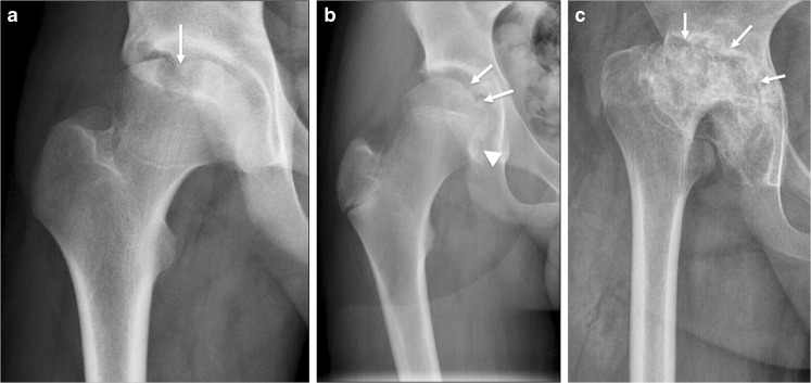
Measuring bone erosions through a variety of destructive changes seen at the hip joint on anteroposterior pelvic radiographs in different children with juvenile idiopathic arthritis. a Radiograph in a 16-year-old boy shows a large bone erosion at the weight-bearing portion of the proximal femoral epiphysis with associated sclerosis (arrow). b Radiograph in a 13-year-old boy shows a subtle bone erosion along the medial femoral neck, just inferior to the physis (arrowhead). Other small erosions are also present at the medial aspect of the proximal femoral epiphysis (arrows). c Radiograph in a 13-year-old girl shows multiple irregularities along both the acetabular roof and femoral head with loss of femoral head height (arrows), in keeping with widespread erosive changes

